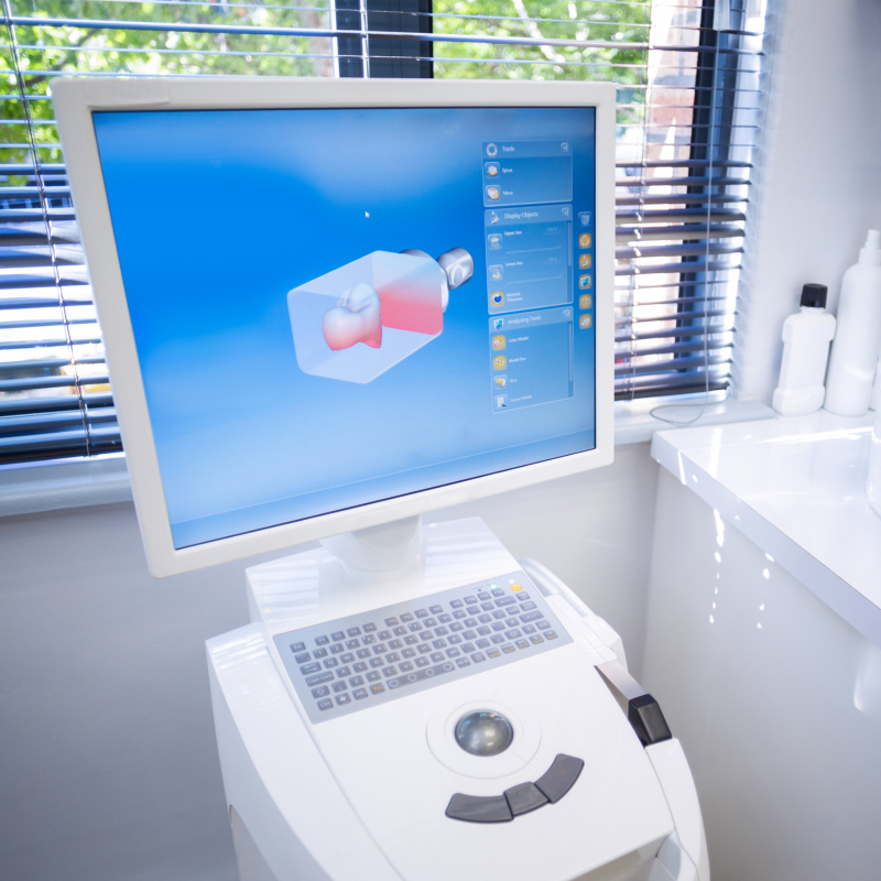
Scanning for a Full Mouth Digital Record with iTero
Efficiency and accuracy are paramount when it comes to restorative cases. The iTero® intraoral scanner is a powerful tool that simplifies the process of scanning and charting for single unit restorations.
The Step-By-Step Process
- Sit behind the patient at the 12 o’clock position with the display on the dominant side for easy access.
- Tap on the scanning sleeve icon to activate the wand after completing the case Rx.
- Wait for 10 seconds for the wand tip to defog before placing it in the patient’s mouth.
- Start scanning by placing the wand tip on the occlusal surface of the terminal molar.
- Scan the entire occlusal surface of the arch, steadily moving the wand towards the anterior.
- Tilt slightly to the lingual at the bicuspid and proceed to the contralateral bicuspid.
- Rotate to the lingual and capture the inner proximal anatomy of the entire lingual surface.
- Rotate to the buccal and use a rocking motion to capture the inner proximal anatomy while moving towards the anterior.
- Move the wand tip towards the opposite terminal tooth and capture the buccal surface using the rocking motion.
- Capture the incisal anatomy by rolling the wand from the lingual surface over the incisal edge to the buccal surface.
- Repeat the same scanning sequence and technique on the contralateral side.
- Use the specified options to move to the next arch and segment.
- Confirm the correct bite by observing the occlusion in the viewfinder.
- Capture three to four teeth in a wave-like motion and repeat on the contralateral side.
- Tap on the view icon to evaluate the digital model in high resolution.
- Add additional scans if necessary or wait for the high-resolution model to be ready.
- Confirm that the scans meet the requirements, including Invisalign-specific criteria.
- Send the case when satisfied with the digital model.
In the rapidly evolving field of dentistry, digital technology has revolutionized various aspects of dental care. One such innovation is the creation of detailed full mouth digital impressions. This technique offers dental professionals a convenient and accurate way to capture the intraoral environment, making the traditional messy and uncomfortable impressions a thing of the past. In this article, we will explore the key steps involved in scanning for a full mouth digital record, equipping dental clinicians with the knowledge to master this art and enhance their clinical workflow.
Step 1: Positioning for Success To ensure optimal access and comfort, begin by sitting behind the patient at the 12 o’clock position, with the display on your dominant side. This arrangement allows easy access to the display touchscreen and the wand, without any unnecessary twisting or turning. By prioritizing ergonomic positioning, you set the stage for a seamless scanning experience.
Step 2: Activating the Wand Once you have completed the necessary case Rx, tap on the scanning sleeve icon at the top of the screen to activate the wand. It’s crucial to allow a 10-second interval after the light turns on, as this defogs the wand tip and ensures clear imaging. This attention to detail sets the foundation for accurate scans.
Step 3: Scanning the Arch Place the wand tip on the occlusal surface of the terminal molar and start scanning the entire occlusal of the arch. Keep the wand flat on the occlusal surface and steadily bring it towards the anterior. As you reach the bicuspid, tilt slightly to the lingual and proceed to the contralateral bicuspid. Capture the inner proximal anatomy of the entire lingual surface by using a twisting motion. Continue scanning, ensuring thorough coverage of the lingual and buccal surfaces, reducing interference from the cheek for a smoother experience.
Step 4: Fine-tuning the Details To ensure comprehensive digital impressions, it’s important to pay attention to the finer details. After completing the lingual scanning, rotate to the buccal surface and employ a rocking motion as you move from posterior to anterior. This technique captures the inner proximal anatomy effectively while minimizing interference from the cheek. Take care to capture the incisal anatomy of the anterior teeth by placing the wand in a position that centers the cuspid and lateral in the viewfinder. Roll the wand from the lingual surface over the incisal edge to the buccal surface, repeating the process on the contralateral side. These scans play a crucial role in joining the lingual and buccal segments accurately.
Step 5: Navigating Arch Segments Once you have completed scanning one arch, it’s time to move to the next segment. You have three options for navigation: tapping on the specified arch on the touchscreen, using the arrow key on the segment indicator box, or activating the touchpad on the wand. Familiarize yourself with these navigation methods to streamline your workflow. If you need to go back to a previous segment, simply swipe from right to left.
Step 6: Capturing the Bite The final segment is capturing the bite, a critical aspect of the full mouth digital record. Before scanning, ensure the correct bite by having the patient open and retract their cheek. In centric occlusion, gently bring the wand tip against the teeth, observing the occlusion in the viewfinder. Utilize a small wave-like motion to capture three to four teeth, then repeat the process on the contralateral side. This step ensures a comprehensive representation of the bite.
Step 7: Evaluating the Digital Model Once you have completed scanning both arches and the bite, it’s time to evaluate the digital model. Tap on the view icon at the top of the touchscreen display to view the high-resolution digital model. Take this opportunity to assess the model’s quality, ensuring that all necessary anatomical structures are included. If needed, you can add additional scans or make adjustments before proceeding.
Step 8: Meeting Invisalign Requirements If the scan is intended for Invisalign treatment, it’s crucial to confirm that the scans meet specific requirements. Check that the digital model includes solid incisal edges, the distal anatomy of the terminal molars, two millimeters of gingival tissue, and accurate interproximal surfaces. Additionally, ensure that the correct bite is captured. Address any areas of missing anatomy indicated in red (in monochrome mode) or purple (in color mode).
Conclusion
Mastering the art of full mouth digital impressions empowers dental professionals with a modern and efficient approach to capturing intraoral information. By following these key steps, clinicians can achieve detailed and accurate digital records, enhancing their clinical workflow and improving patient experiences. Embracing digital technology in dentistry opens up new possibilities, revolutionizing the way clinicians provide care and ensuring optimal outcomes for their patients. Stay updated with the latest advancements in digital dentistry and embrace the transformative power of technology.


